
by holistic.eye | Jul 28, 2015 | Sclera Markings
A ‘Bitot’s Spot’ Sclerology marking is a sign of vitamin A deficiency and appears medially and laterally toward the border of the iris as a superficial, opaque, foamy patch in the conjunctiva. Common signs & symptoms of vitamin A deficiency are...

by holistic.eye | Jul 28, 2015 | Sclera Markings
Cystine is a semi-essential amino acid, the most important of the 3 amino acids (glycine, glutamate, cystine), that are required by the body for the production of glutathione. Cystine deposits are accumulations of crystalline cystine in the sclera and cornea. Seen...

by holistic.eye | Jul 27, 2015 | Sclera Markings
This sclera image shows, at this stage relatively faintly, Sclerology marking ‘boxing’ in the lymph/breast area. This marking presents as its name suggests, shaped like open-ended boxes. It represents local tissue hardening, and can suggest possible nodes...
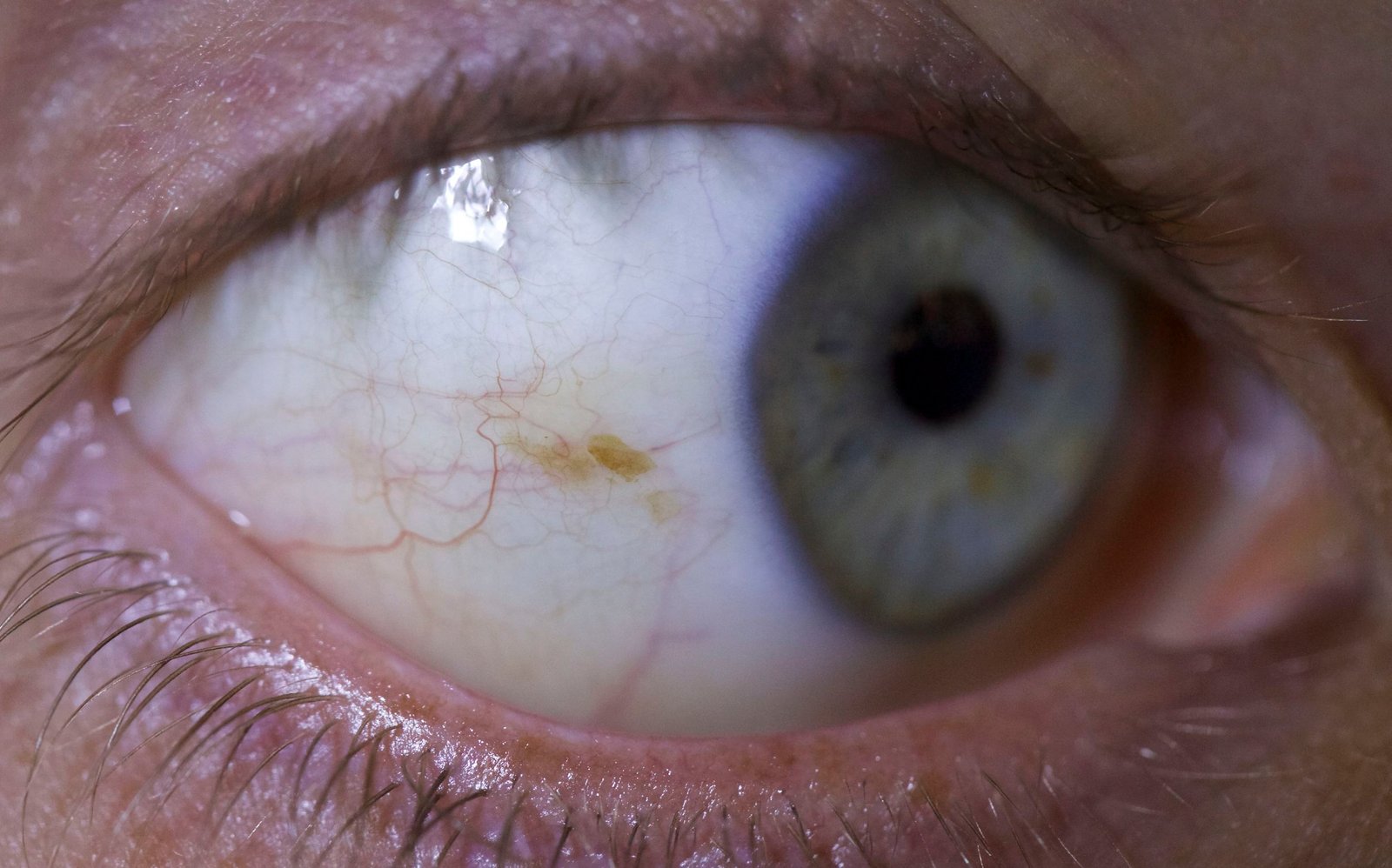
by holistic.eye | Jun 25, 2015 | Sclera Markings
This right lateral sclera image shows melanin pigment, the brownish coloured ‘splotch’ you can see in the sclera. This Sclerology marking is common as a pigmentation spot in a person with darker skin and brown/hazel eyes, representing a genetic...

by holistic.eye | May 25, 2015 | Recipes
Chia seeds are a very rich source of calcium, magnesium, phosphorous and manganese, antioxidants, fibre, omega-3 fatty acids and HDL (good cholesterol). They also provide good satiety due to the fat and protein content, help to stabilise blood sugar levels and...
